
What Is a Sialogram? Uses, Procedure, Results, Risks, and Modern Alternatives


What Is a Sialogram?
Dry mouth, swelling under your jaw, or pain while eating may be signs of a problem with your salivary glands rather than your teeth.
A sialogram is a specialized imaging test that checks the salivary glands and ducts to find the cause of these symptoms. It helps identify blocked ducts, salivary stones, narrowing, or other conditions that affect the normal flow of saliva.
Your dentist will evaluate your oral health to see whether your symptoms are caused by a dental condition or another underlying issue. If a salivary gland condition is suspected, you may be referred to a specialist for a sialogram.
The results help determine the cause of your symptoms and guide the next steps in your treatment.
Understanding Your Salivary Glands
Your salivary glands make saliva, which helps you chew, swallow, digest food, and protect your teeth by washing away food particles and bacteria.
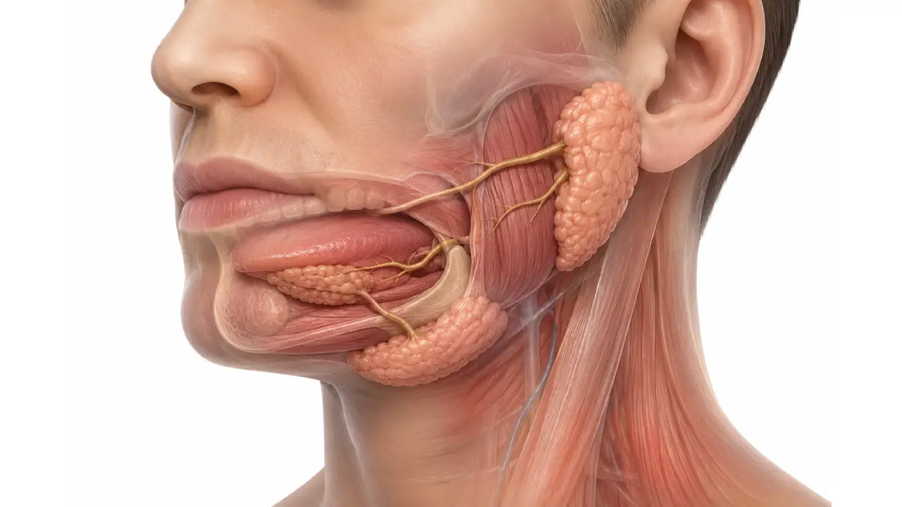
There are three major pairs of salivary glands:
Parotid Glands
Located in front of your ears, these are the largest salivary glands. When you eat, these glands release saliva through small ducts into your mouth.
Submandibular Glands
These glands sit beneath your lower jaw and produce most of your saliva throughout the day. They are also the most common place for salivary stones to develop.
Sublingual Glands
Located under your tongue, these glands release saliva through several small ducts to help keep your mouth moist.
How Saliva Flows
Saliva travels from the glands to your mouth through small tubes called salivary ducts. If one of these ducts becomes blocked or narrowed, saliva cannot flow properly. This can lead to symptoms such as:
- Swelling under the jaw or near the ears
- Pain while eating
- Dry mouth
- Repeated salivary gland infections
Because these symptoms can feel similar to a tooth or jaw problem, your dentist will examine your mouth to determine whether the cause is dental or related to your salivary glands.
When Might a Dentist Recommend a Sialogram?
First, your dentist will examine your teeth, gums, and jaw to rule out common dental problems. If a salivary gland problem is suspected, you may be referred for a sialogram.
Your dentist may recommend this test if you have:
- Swelling under the jaw or near the ears, especially during or after meals
- Ongoing dry mouth
- Pain while eating or swallowing
- Repeated salivary gland infections
- Thick or reduced saliva
- Difficulty swallowing due to reduced saliva
- A suspected salivary stone or blocked duct
- Unexplained swelling in the face or jaw
Swelling that appears during meals and improves afterward is a common sign of a blocked salivary duct. A sialogram can help identify the cause and guide the right treatment.
Conditions a Sialogram Can Help Diagnose
Chronic Salivary Gland Inflammation (Chronic Sialadenitis)
Some patients develop repeated infections because saliva cannot drain properly. When saliva becomes trapped inside a blocked or narrowed duct, bacteria may multiply more easily, increasing the risk of infection.
Symptoms may include:
- Swelling of the affected gland
- Redness around the opening of the duct
- Fever
- Pus draining into the mouth
- Difficulty opening the mouth
- Pain while chewing
Although infections are usually diagnosed through a clinical examination, imaging may be recommended if symptoms continue to return despite treatment.
Sjogren's Syndrome
Sjogren’s syndrome is an autoimmune condition in which the body’s immune system mistakenly attacks the glands responsible for producing saliva and tears.
People with Sjogren’s syndrome commonly experience:
- Persistent dry mouth
- Dry eyes
- Difficulty swallowing dry foods
- Increased risk of tooth decay
- Frequent oral infections
A sialogram may reveal changes in the salivary ducts caused by long-term gland damage. However, diagnosing Sjogren’s syndrome usually requires additional blood tests, medical history, and evaluation by a physician or rheumatologist.
Structural Abnormalities
Some people are born with unusually shaped salivary ducts, while others develop structural changes after surgery, trauma, or repeated inflammation.
These abnormalities may interfere with normal saliva flow and increase the likelihood of chronic swelling or infections.
A sialogram helps specialists visualize the duct system and determine whether the abnormality contributes to the patient’s symptoms.
What Happens Before a Sialogram?
Most patients require very little preparation before a sialogram. Your healthcare provider will review your medical history and explain the procedure before it begins.
Before the appointment, tell your healthcare provider if you:
- Have allergies to iodine or contrast dye
- Are pregnant or think you may be pregnant
- Are breastfeeding
- Have an active salivary gland infection
- Take blood-thinning medications
- Have recently undergone surgery involving the mouth or jaw
Depending on your situation, your healthcare provider may recommend delaying the procedure until an active infection has been treated.
Before the imaging begins, you may also be asked to:
- Remove removable dentures or oral appliances
- Remove earrings or facial jewelry near the imaging area
- Rinse your mouth to reduce bacteria around the duct opening
Most patients do not need sedation and can return home shortly after the examination.
What Happens During a Sialogram?
Understanding each step of the procedure can help reduce anxiety and make the experience more comfortable.
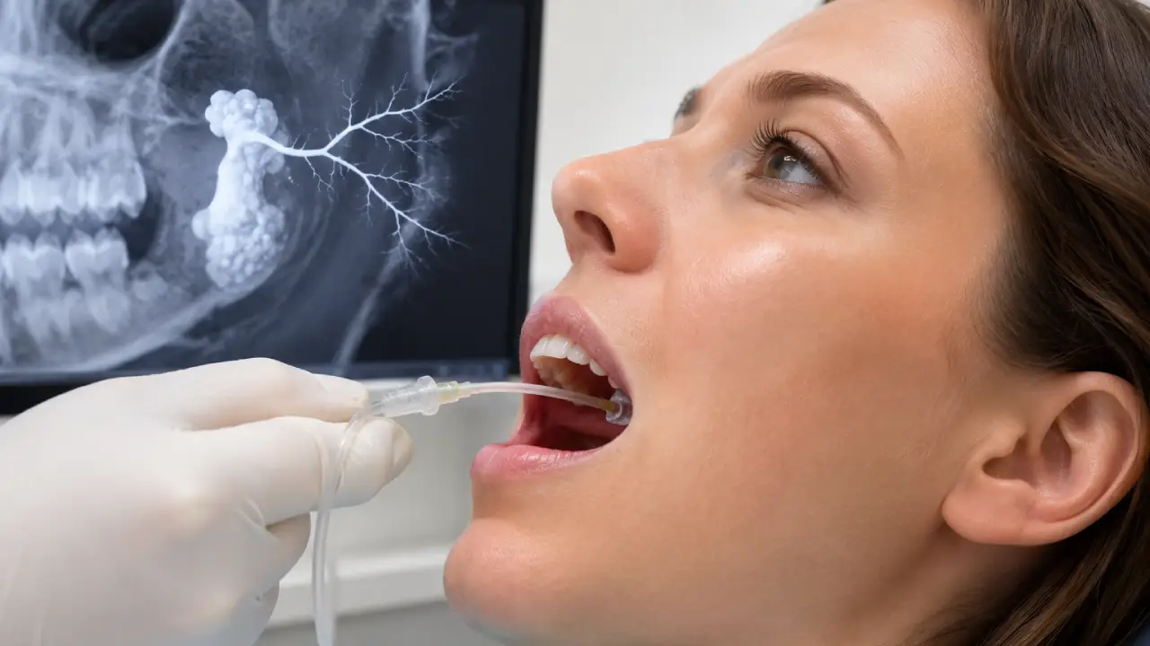
Step 1: Locating the Salivary Duct
The healthcare provider first identifies the tiny opening of the salivary duct inside your mouth. This opening is carefully cleaned before the procedure begins.
Step 2: Inserting a Small Catheter
A very thin catheter is gently inserted into the duct opening.
Although this sounds uncomfortable, most patients describe the sensation as mild pressure rather than pain.
If necessary, a local anesthetic may be used to improve comfort.
Step 3: Injecting the Contrast Material
A small amount of contrast solution is slowly injected into the duct.
The contrast outlines the duct system, making it visible on X-ray images.
Some patients notice:
- Mild pressure
- A feeling of fullness
- A temporary unusual taste
These sensations typically disappear within a few minutes.
Step 4: Taking X-ray Images
Several X-ray images are taken from different angles while the contrast remains inside the duct.
These images help the radiologist evaluate:
- Saliva flow
- Duct shape
- Blockages
- Narrowed areas
- Salivary stones
- Structural abnormalities
You may be asked to change head position slightly while the images are taken.
Step 5: Stimulating Saliva Flow Salivary Duct
Once the initial images are complete, you may be given a sour substance, such as lemon juice or a citric acid preparation.
This stimulates saliva production and helps empty the contrast material from the gland.
Additional images may then be taken to observe how effectively saliva clears from the duct system.
How Long Does a Sialogram Take?
Although appointment times vary, the actual imaging procedure usually takes between 30 and 45 minutes.
Most patients can return to normal daily activities immediately afterward unless instructed otherwise by their healthcare provider.
Does a Sialogram Hurt?
One of the most common questions patients ask is whether a sialogram is painful.
For most people, the procedure causes only mild and temporary discomfort.
You may experience:
- Slight pressure during catheter placement
- Mild fullness while the contrast is injected
- A temporary metallic or bitter taste
- Minor tenderness around the gland afterward
Significant pain is uncommon.
If you experience severe discomfort during the procedure, let the healthcare provider know immediately. They can often adjust the catheter or provide additional measures to improve your comfort.
Some patients notice mild swelling for a few hours after the examination. Drinking water, gently massaging the gland if advised, and allowing saliva to flow naturally often help relieve this sensation.
Understanding Your Sialogram Results
A radiologist carefully reviews the images before sending a report to the healthcare provider who ordered the examination.
Your dentist or specialist will then discuss the findings and explain whether additional treatment or testing is necessary.
Normal Results
A normal sialogram shows:
- Smooth salivary ducts
- No evidence of obstruction
- Normal saliva drainage
- Healthy gland appearance
If the results are normal, your healthcare provider may investigate other possible causes of your symptoms.
Abnormal Results
Depending on the images, a sialogram may identify:
Salivary Stones
Mineral deposits blocking saliva flow.
Narrowed Ducts
Areas where inflammation or scarring has reduced the duct diameter.
Chronic Inflammation
Irregular duct patterns suggesting long-standing gland disease.
Leakage of Contrast
Rarely, contrast may escape outside the duct, indicating injury or damage.
Structural Changes
Unusual duct branching or abnormalities affecting saliva drainage.
Sometimes the findings are enough to guide treatment. In other situations, additional imaging such as ultrasound, MRI, or CT may be recommended for a more complete evaluation.
Risks and Safety Considerations
A sialogram is generally considered a safe diagnostic procedure when performed by trained healthcare professionals.
Like any medical test, however, it carries some potential risks.
Mild Radiation Exposure
Because the procedure uses X-rays, patients are exposed to a small amount of radiation.
The exposure is relatively low, and healthcare providers use the minimum amount necessary to obtain diagnostic images.
If you are pregnant or think you may be pregnant, inform your healthcare provider before the examination so alternative imaging options can be considered if appropriate.
Temporary Swelling or Discomfort
Some patients experience mild tenderness or swelling after the procedure.
These symptoms are usually temporary and improve within a day.
Allergic Reaction to Contrast Material
Although uncommon, some individuals may experience an allergic reaction to the contrast solution.
For this reason, your healthcare provider will ask about previous reactions to iodine or contrast agents before the procedure begins.
Infection
Because a catheter is placed into the salivary duct, there is a very small risk of introducing bacteria.
Proper sterile technique keeps this risk extremely low.
Contact your healthcare provider if you develop:
- Fever
- Increasing swelling
- Severe pain
- Pus draining from the duct
- Persistent bleeding
These symptoms may require prompt medical evaluation.
Modern Alternatives to a Sialogram
Advances in medical imaging have introduced several alternatives that may be recommended depending on your symptoms and the suspected condition.
| Imaging Test | Best Used For | Contrast Dye | Radiation |
| Ultrasound | Detecting salivary stones and gland swelling | No | No |
| CBCT | Calcified stones and surrounding bone structures | No | Low |
| MRI | Soft tissue evaluation and tumors | Usually No | No |
| CT Scan | Complex infections and deep tissue involvement | Sometimes | Yes |
| Sialogram | Detailed salivary duct imaging | Yes | Low |
| Sialendoscopy | Diagnosing and treating duct blockages | No | Minimal |

What Happens After Diagnosis?
Your treatment will depend on what the sialogram shows. Your healthcare provider will review the results along with your symptoms and recommend the most appropriate treatment plan.
If a Salivary Stone Is Found
If a salivary stone is causing the blockage, treatment depends on its size and location. Small stones may pass naturally with increased hydration and saliva stimulation, while larger stones may need to be removed using a minimally invasive procedure called sialendoscopy. Surgery is only recommended in rare cases when other treatments are not successful.
If an Infection Is Found
When a salivary gland infection is diagnosed, treatment usually includes antibiotics to clear the infection and medication to relieve pain and swelling. Your healthcare provider may also recommend drinking plenty of water and using warm compresses to help improve saliva flow. Most patients begin to feel better within a few days of starting treatment.
If the Salivary Duct Is Narrowed
A narrowed salivary duct can reduce the flow of saliva and cause repeated swelling or discomfort. Depending on the severity, your healthcare provider may recommend monitoring the condition, prescribing medication to reduce inflammation, or performing a sialendoscopy to widen the duct. Surgery is only considered if other treatments are not effective.
If an Autoimmune Condition Is Suspected
If the results suggest an autoimmune condition such as Sjogren’s syndrome, additional tests will usually be needed to confirm the diagnosis. Your healthcare provider may recommend blood tests, saliva flow tests, or refer you to a specialist for further evaluation. Treatment focuses on managing symptoms, improving saliva production, and protecting your oral health.
If a Growth or Tumor Is Found
If a growth is identified, further evaluation is needed to determine whether it is benign or requires additional treatment. Your healthcare provider may recommend imaging tests such as an ultrasound, MRI, or CT scan, and in some cases a biopsy. Although many salivary gland tumors are non-cancerous, early diagnosis helps ensure the most appropriate treatment.
Frequently Asked Questions
1. What is a sialogram used for?
A sialogram helps healthcare providers evaluate the salivary glands and ducts for blockages, salivary stones, chronic inflammation, narrowing, and other conditions that interfere with normal saliva flow.
2. Is a sialogram painful?
Most patients experience only mild discomfort. You may notice slight pressure during catheter placement or a temporary feeling of fullness when the contrast material is injected, but significant pain is uncommon.
3. How long does a sialogram take?
The procedure typically takes 30 to 45 minutes, although appointment times may vary depending on the complexity of the examination.
4. Can I eat after a sialogram?
In most cases, yes. Unless your healthcare provider gives different instructions, you can usually return to your normal eating and drinking routine shortly after the procedure.
5. Who performs a sialogram?
A sialogram is usually performed by a radiologist or another trained healthcare professional at a hospital or specialized imaging center. General dental offices typically refer patients for this examination rather than performing it on-site.
6. Can a sialogram detect cancer?
A sialogram may reveal abnormalities that require further investigation, but it is not designed to diagnose cancer. If a suspicious mass is identified, additional imaging or a biopsy may be recommended.
7. Are there alternatives to a sialogram?
Yes. Depending on your symptoms, your healthcare provider may recommend an ultrasound, CBCT scan, MRI, CT scan, or sialendoscopy instead of a sialogram.

Conclusion
A sialogram is a useful imaging test for finding problems that affect the salivary glands and ducts, such as blockages, salivary stones, or chronic inflammation. It helps healthcare providers identify the cause of your symptoms and recommend the right treatment.
If you have ongoing dry mouth, swelling under your jaw, pain while eating, or repeated salivary gland infections, it’s important to have these symptoms evaluated. A dental examination can help determine whether the problem is related to your teeth or your salivary glands.
At AL Dental Studio, we carefully assess your symptoms and perform a thorough dental examination. If a salivary gland condition is suspected, we’ll refer you to the appropriate specialist for advanced imaging and treatment. Early diagnosis can help prevent complications and ensure you receive the care you need.
Dental Experts, You Can Trust
Medically Reviewed. Last updated on July 29, 2026.
Learn more about our editorial standards.

Dr. Alexander Heifitz (Author)
Dr. Alexander Heifitz is the founder of AL Dental Studio in NYC, where he combines advanced dental expertise with a patient-first approach. He specializes in cosmetic and restorative treatments such as dental implants, veneers, Invisalign, and smile makeovers, helping New Yorkers achieve both oral health and confidence.

Book An Appointment
Looking for a reliable dentist in Upper East NYC? Whether you need a routine cleaning, urgent care, or a full smile transformation, we’ve got you covered. We accept most PPO insurance plans and offer flexible scheduling.
+1 (212) 430-3888
Call for appointment
Walk-ins Welcome / Same-Day Appointments Available

Related Blogs

Mini Dental Implants NYC: Cost, Benefits & Fast Recovery Guide
Written by Dr. Alexander Heifitz, Founder of AL Dental Studio | Medically Reviewed. Last updated on February 20, 2026.
Read More
Zygomatic Dental Implants: Are They Right for You in NYC?
Written by Dr. Alexander Heifitz, Founder of AL Dental Studio | Medically Reviewed. Last updated on February 19, 2026.
Read More